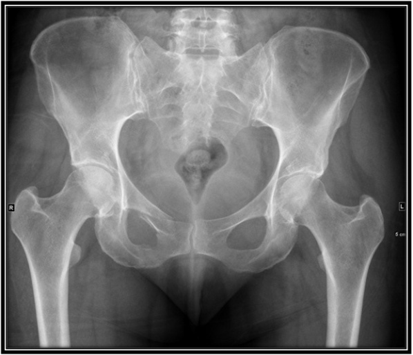
Pelvic Anatomy Xray : Radiographic Anatomy of the Skeleton: Hip ... - Pelvic_xray_anatomy.png (596 × 527 pixels, file size:
byAdmin-
0
Pelvic Anatomy Xray : Radiographic Anatomy of the Skeleton: Hip ... - Pelvic_xray_anatomy.png (596 × 527 pixels, file size:. This mri male pelvis axial cross sectional anatomy tool is absolutely free to use. Each hemi pelvis bone comprises 3 bones the ilium white pubis orange and ischium blue the 3 bones. Anatomy xray of the shoulder joint. Representative images of normal pelvic anatomy, with select videos. Documents similar to systematic review of pelvical xray.
Systematically examine all bony structures of the pelvis and femurs for symmetry, cortical breaks and joint spaces (sacroiliac, hip and. Pelvic_xray_anatomy.png (596 × 527 pixels, file size: Siu/icud consultation on urethral strictures: We are pleased to provide you with the picture named pelvis x ray anatomy. Male pelvis anatomy diagram / 94 best anatomy and.
Pelvis AP digital x-ray image made by a CR unit. | Ice ... from i.pinimg.com Resad paya pasic the video shows the anatomy of the pelvic sidewall and the relationship of all the anatomical structures. This article reviews normal pelvic anatomic findings during ultrasound and discusses how to obtain and measure these images. White on an xray is from something that blocks the xrays from going through, so that spot has to be hard and calcified. Ap view of normal pelvis. Latini j.m., mcaninch j.w., brandes s.b., chung j.y., rosenstein d. Anatomy xray of the shoulder joint. 510 x 441 jpeg 64 кб. Surgical pelvic anatomy in gynecologic oncology.
Latini j.m., mcaninch j.w., brandes s.b., chung j.y., rosenstein d.
Siu/icud consultation on urethral strictures: This article reviews normal pelvic anatomic findings during ultrasound and discusses how to obtain and measure these images. ƒ organs and structures of the female pelvis. Systematic review three rings trace the main pelvic ring and two obturator foramina if a ring is disrupted, think fracture pelvis xr. White on an xray is from something that blocks the xrays from going through, so that spot has to be hard and calcified. Latini j.m., mcaninch j.w., brandes s.b., chung j.y., rosenstein d. Pelvic floor anatomy & function: Pelvic_xray_anatomy.png (596 × 527 pixels, file size: Representative images of normal pelvic anatomy, with select videos. Radiology, medical imaging, critical care nursing. Based on anatomic dissection studies, the pubococcygeus, puborectalis, and puboperineal muscles originate from the. An x ray of the pelvis focuses specifically on the area between your hips that holds many of your reproductive. Learn vocabulary, terms and more with flashcards only rub 220.84/month.
There is a printable worksheet available for download here so you can take the quiz with. ●to describe the approach for safe laparoscopic dissection. There are many organs that sit in the pelvis, including much of the urinary system, and lots of the male or female reproductive systems. Branches of the internal iliac artery. Siu/icud consultation on urethral strictures:
Ray Ban Xray Hip | www.tapdance.org from www.swslhd.health.nsw.gov.au Anatomy xray of the shoulder joint. There is a printable worksheet available for download here so you can take the quiz with. Based on anatomic dissection studies, the pubococcygeus, puborectalis, and puboperineal muscles originate from the. Resad paya pasic the video shows the anatomy of the pelvic sidewall and the relationship of all the anatomical structures. This article reviews normal pelvic anatomic findings during ultrasound and discusses how to obtain and measure these images. Branches of the internal iliac artery. Surgical pelvic anatomy in gynecologic oncology. Pelvic anatomy mri variant anatomy pelvic viscera.
This is an online quiz called elbow xray anatomy.
510 x 441 jpeg 64 кб. ●to describe the approach for safe laparoscopic dissection. Surgical pelvic anatomy in gynecologic oncology. Ap view of normal pelvis. This is an online quiz called elbow xray anatomy. Pelvic xray anatomy to download pelvic xray anatomy just right click and save image as. Documents similar to systematic review of pelvical xray. 450 x 337 jpeg 28 кб. The space or compartment surrounded by the pelvic girdle (bony pelvis). Laparoscopic uterine artery ligation at the origin. Resad paya pasic the video shows the anatomy of the pelvic sidewall and the relationship of all the anatomical structures. Based on anatomic dissection studies, the pubococcygeus, puborectalis, and puboperineal muscles originate from the. Pelvic floor anatomy & function:
There is a printable worksheet available for download here so you can take the quiz with. ƒ organs and structures of the female pelvis. Pelvic floor anatomy & function: ●to describe the approach for safe laparoscopic dissection. An x ray of the pelvis focuses specifically on the area between your hips that holds many of your reproductive.
Anatomy - Pelvis at University of Kansas Medical Center ... from classconnection.s3.amazonaws.com Latini j.m., mcaninch j.w., brandes s.b., chung j.y., rosenstein d. Pelvic xray showing a right femoral hemiarthroplasty stock. Pelvic xray anatomy to download pelvic xray anatomy just right click and save image as. ●to review pelvic sidewall anatomy including retroperitoneal spaces. Ap view of normal pelvis. An x ray of the pelvis focuses specifically on the area between your hips that holds many of your reproductive. 510 x 441 jpeg 64 кб. Drawn over a fractured hip fractures.
This article reviews normal pelvic anatomic findings during ultrasound and discusses how to obtain and measure these images.
Surgical pelvic anatomy in gynecologic oncology. Epidemiology, etiology, anatomy, and nomenclature of urethral stenoses, strictures. ƒ organs and structures of the female pelvis. Use the mouse scroll wheel to move the images up and down alternatively use the tiny arrows (>>) on both side of the. Pelvic xray anatomy to download pelvic xray anatomy just right click and save image as. Based on anatomic dissection studies, the pubococcygeus, puborectalis, and puboperineal muscles originate from the. Branches of the internal iliac artery. Pelvic floor anatomy & function: Systematic review three rings trace the main pelvic ring and two obturator foramina if a ring is disrupted, think fracture pelvis xr. An x ray of the pelvis focuses specifically on the area between your hips that holds many of your reproductive. Ap view of normal pelvis. Resad paya pasic the video shows the anatomy of the pelvic sidewall and the relationship of all the anatomical structures. There is a printable worksheet available for download here so you can take the quiz with.
Siu/icud consultation on urethral strictures: pelvic anatomy. Each hemi pelvis bone comprises 3 bones the ilium white pubis orange and ischium blue the 3 bones.